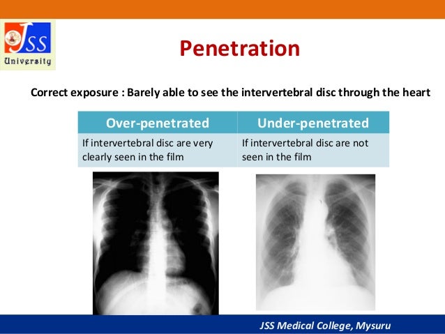

Changes in the quantity and the quality of radiation exposure to a film-screen IR affect the amount of density and contrast visible on the processed radiograph.

Radiographic film acquires the latent image and needs to be chemically processed before the image can be displayed. However, the amount of exposure to the digital IR needs to be carefully selected, as with film-screen IRs, to produce a quality image with the least amount of exposure to the patient. The level of brightness and contrast can be altered during computer processing and image display. Digital IRs separate acquisition from processing and image display their response to changes in radiation exposure does not affect the amount of brightness displayed on the image. Knowledge of how these factors affect the image individually and in combination assists the radiographer to produce a radiographic image with the amount of information desired for a diagnosis.īecause various types of IRs respond differently to the radiation exiting the patient, these differences are noted throughout this chapter. Radiographers have the responsibility of selecting the combination of exposure factors to produce a good-quality image. This chapter focuses on exposure techniques and the use of accessory devices and their effect on the radiation reaching the image receptor (IR) and the image produced. The characteristics evaluated for image quality are density or brightness, contrast, recorded detail or spatial resolution, distortion, and noise. Chapter 3 emphasized that a good-quality radiographic image accurately represents the anatomic area of interest. Milliamperage and time affect the quantity of radiation produced, and kilovoltage affects both the quantity and the quality. In Chapter 2, variables that affect both the quantity and the quality of the x-ray beam were presented. State exposure technique modifications for the following considerations: body habitus, pediatric patients, projections and positions, soft tissue, casts and splints, and pathologic conditions. Identify the exposure factors that can affect patient radiation exposure.ġ7. Recognize patient factors that may affect IR exposure.ġ6. Calculate changes in mAs when adding or removing a grid.ġ5. Describe the use of grids and beam restriction and their effect on IR exposure and image quality.ġ4. Calculate the magnification factor, and determine image and object size.ġ3. Calculate changes in mAs for changes in source-to-image receptor distance.ġ2. Recognize the factors that affect recorded detail and distortion.ġ1. Compare the effect of changes in kVp on digital and film-screen images.ġ0. Calculate changes in kVp to change or maintain exposure to the IR.ĩ. Explain how kilovoltage peak (kVp) affects radiation production and IR exposure.Ĩ.
#UNDER EXPOSURE X RAY HOW TO#
Recognize how to correct exposure factors for a density error.ħ. Compare the effect of changes in milliamperage (mA) and exposure time on digital and film-screen images.Ħ. Calculate changes in milliamperage and exposure time to change or maintain exposure to the IR.ĥ. Explain the relationship between milliamperage and exposure time with radiation production and image receptor (IR) exposure.Ĥ. State all the important relationships in this chapter.ģ. Define all the key terms in this chapter.Ģ. After completing this chapter, the reader will be able to perform the following:ġ.


 0 kommentar(er)
